Alert Box
Notification Box
Electron Microscopes
Microscopes
Image

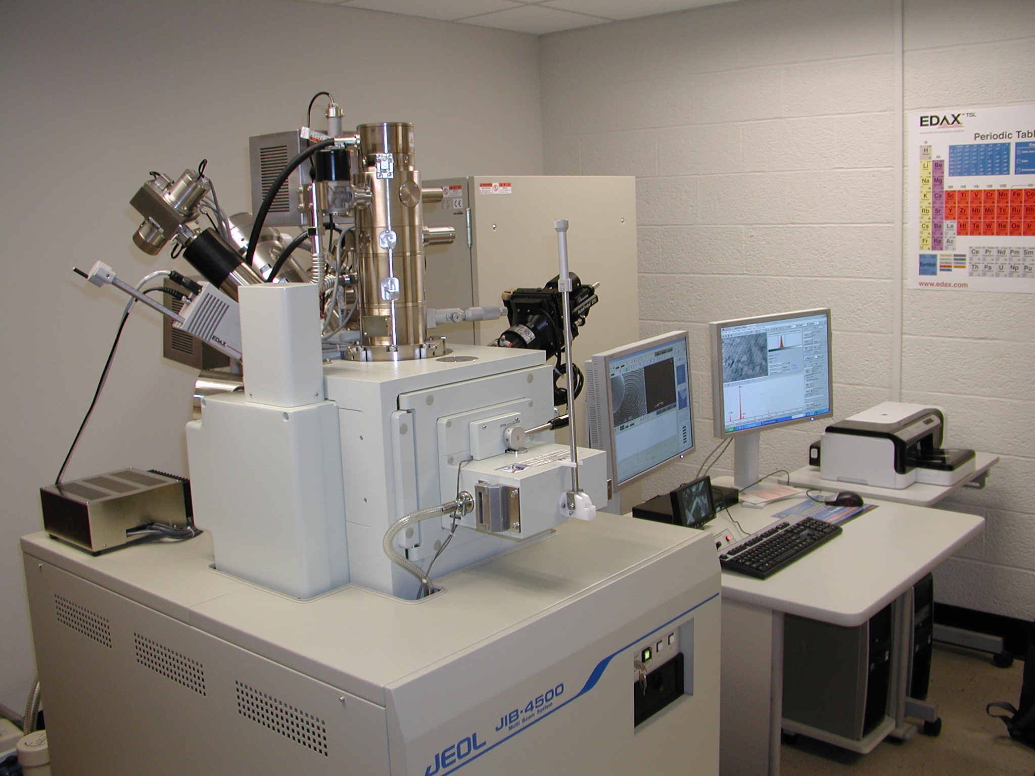
JEOL Jib-4500 Multi beam system
- LaB6 emitter; Acceleration voltage 0.3 to 30,000 V
- Focused Ion Beam (FIB) resolution :5 nm at 30 keV
- Scanning Electron Microscope (SEM) resolution : 2.5 nm at 30 keV
- Pressure range in low-vacuum mode: 1,10 to 100 Pa
- Gas Injection System (GIS) – allows ion-beam deposition of C and W
- X-Ray Energy-Dispersive Spectrometer (EDS) – EDAX APOLLO XV
- EDS resolution, 128 eV measured at MnK, 100,000CPS
- Omniprobe, OMP-AUTOPROBE 200.1 Nanomanipulator
- Remote access capabilities
Image

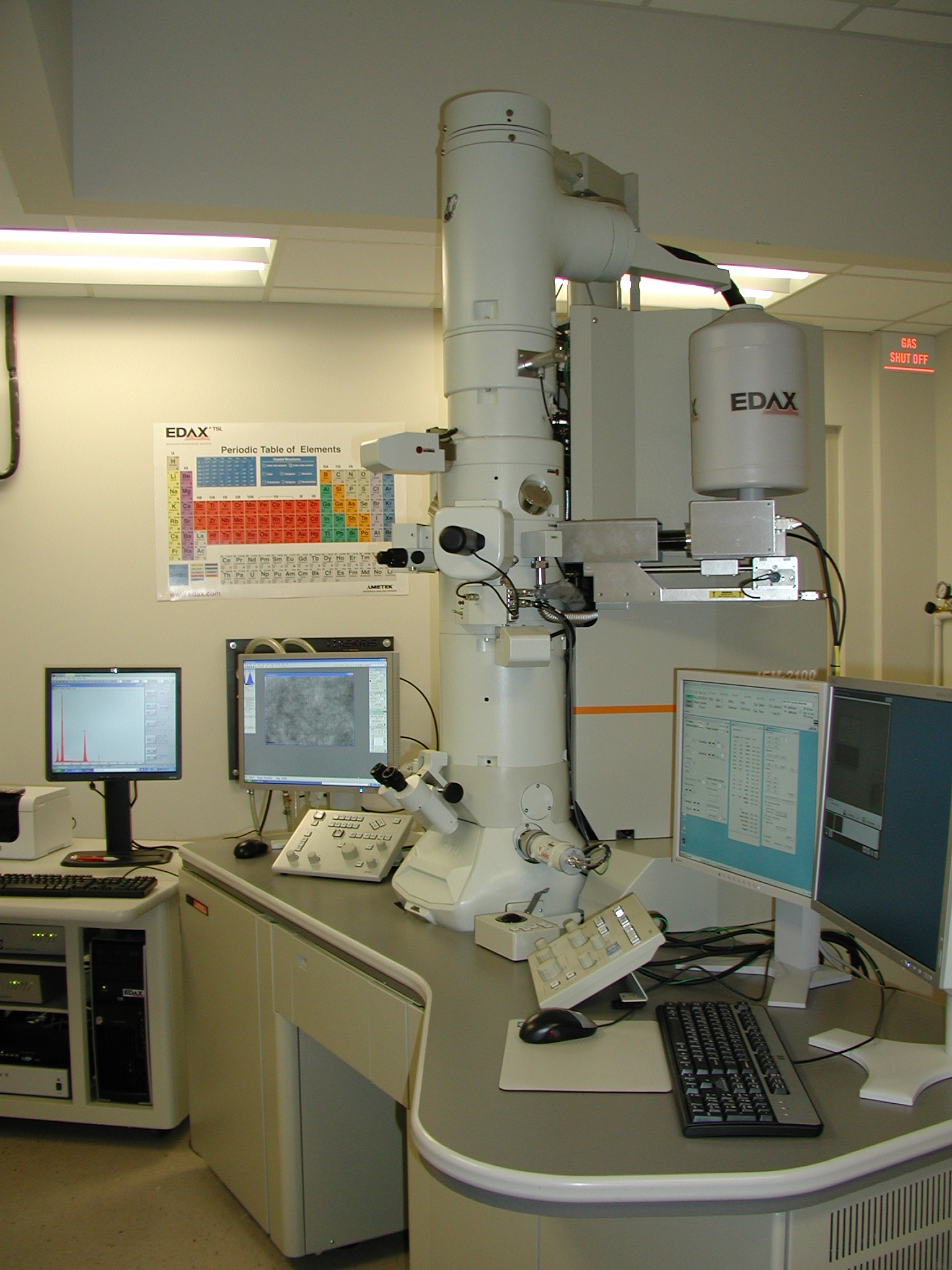
JEOL 2100 Scanning/transmission electron microscope
Instrument features and capabilities:
- Accelerating Voltage up to 200,000 Volts
- Magnification Range ´200 to ´1,500,000
- ~0.20 nm Point-to-Point Resolution; 0.14 nm Lattice Resolution
-
Energy Dispersive X-Ray Detector (with Light Element Detection Capability) for Quantitative Analysis
-
High Resolution (~0.30 nm) Chemical Profile Mapping in Scanning Mode
-
Digital Camera System Enabling Capture of Both High Resolution Images and Electron Diffraction Patterns
-
Remote Access Capability via Knobset Interface Box or Software Only
Image

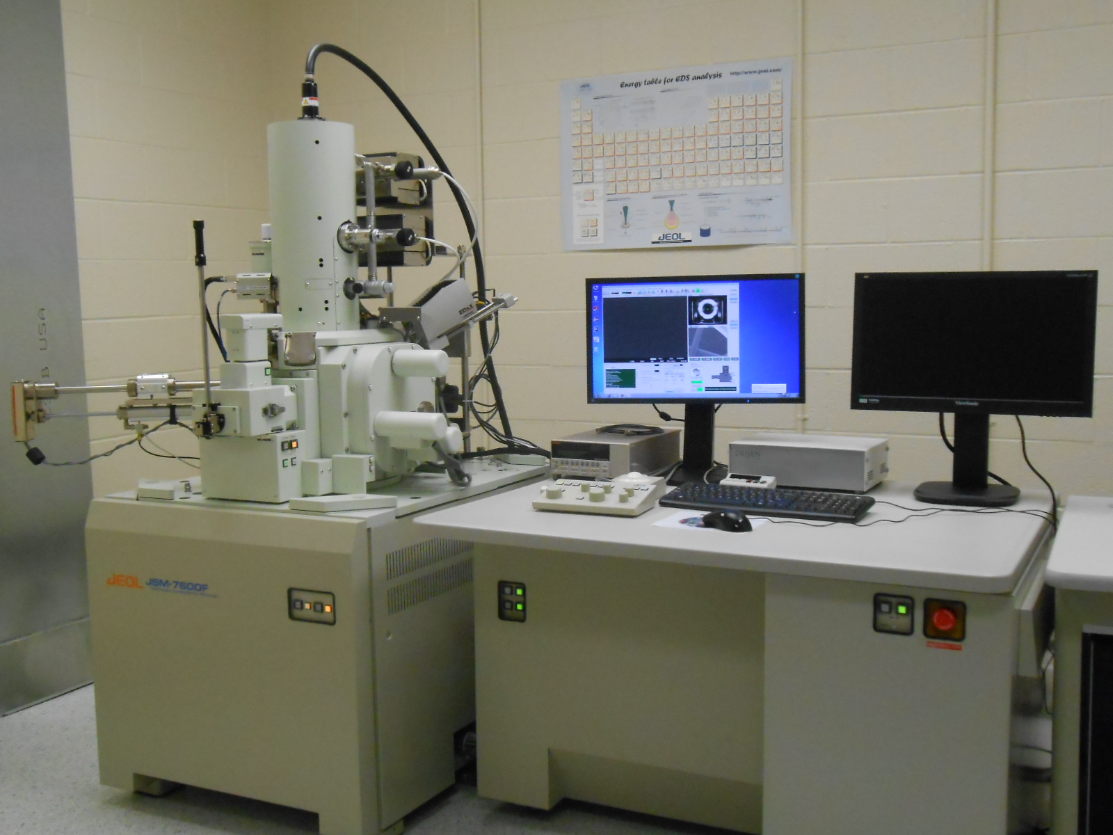
JEOL JSM-7600F Scanning Electron Microscope
- SEI resolution: 1.5 nm (1 kV) in GB mode, 1.0 nm (15 kV)
- Magnification: 25 to 1,000,000x
- Accelerating voltage: 0.1 to 30 kV
- Beam current : 1 pA to 200 nA at 15 kV
- Detector: Upper and lower detectors
- Scanning Transmission Electron Microscopy (STEM) mode
- BS, EDS, EBSD, NPGS systems available
Image

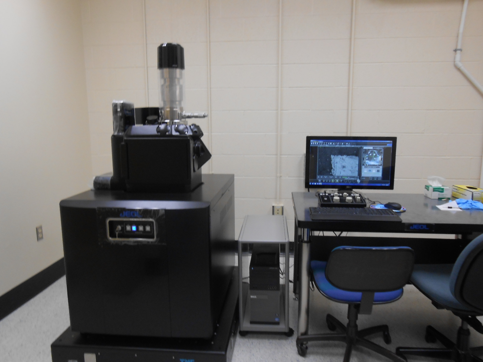
JEOL JSM-IT300LV Variable Pressure SEM
- Extended vacuum range from 10 Pa to 650 Pa
- Accelerating Voltage: 300v to 30kV
- Magnification: 5x to 300,000x
- Resolution: high vacuum model 3.0nm at 30kV; low vacuum model 4.0 nm at 30kV.
- Large analytical chamber and specimen stage can support samples as large as 300mm in diameter
- New intuitive multi-touch software interface
- High resolution imaging with tungsten source
- Mechanically eucentric 5-axis motorized stage with asynchronous movement
- Navigation from a color image built in
- Multiple Live Imaging (including Picture in Picture and signal mixing)
- Video capture built in (AVI format)
Image

To schedule, please click here.
